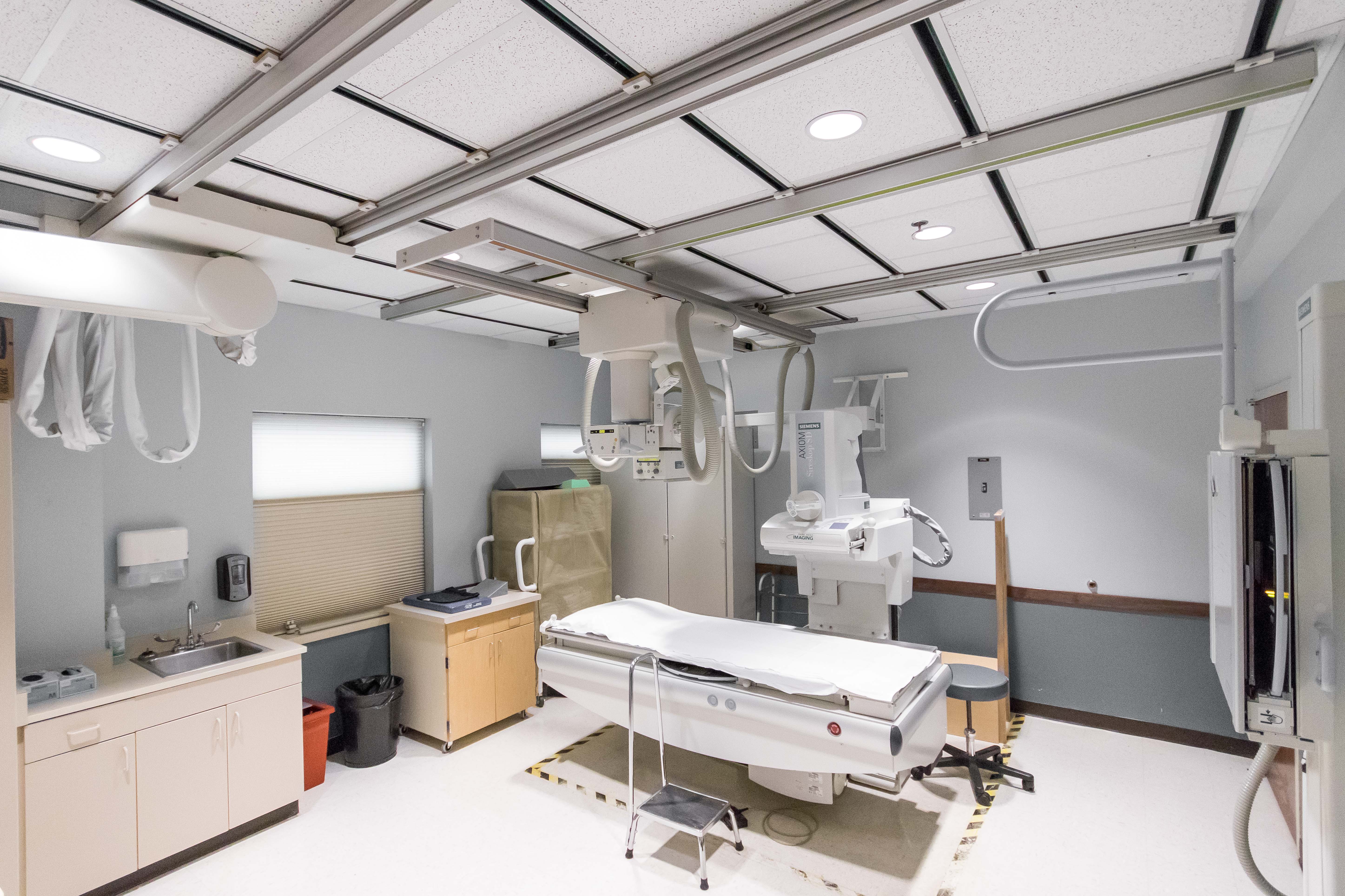
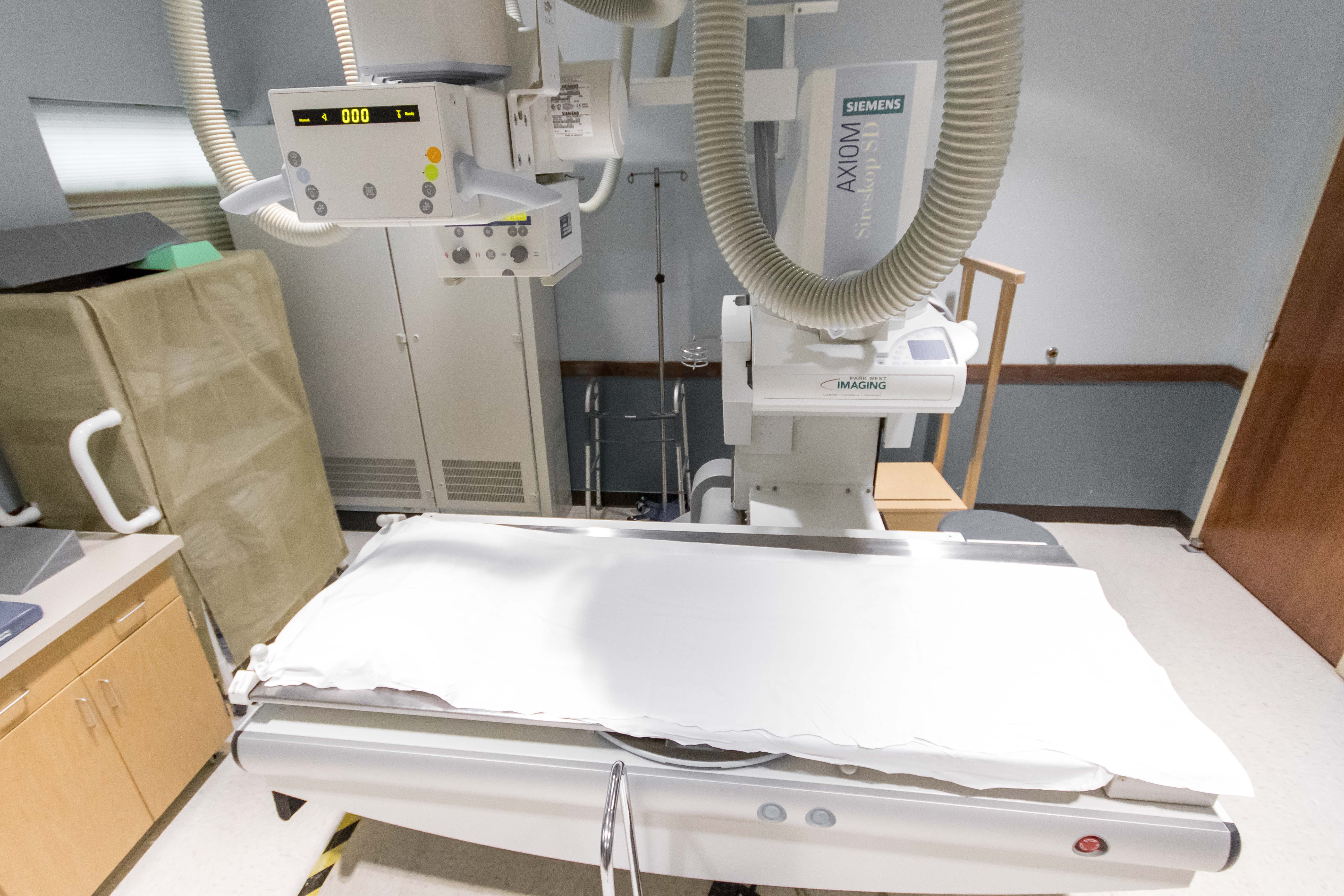
X-ray is high energy electromagnetic radiation that produce 2-dimensional images of the body. Most commonly, X-ray images are used to evaluate bone but can also be used to determine other abnormalities within the human body.
Fluoroscopy is another form of "x-ray" but instead of taking still images of a body structure, fluoro uses a continuous x-ray beam to produce live "movie-like" images. Often, fluoroscopy is used in conjunction with the administration of a contrast agent in order to evaluate a moving structure such as the esophagus or bowel. Fluoroscopy can also be used for exams such as an arthrogram, in which a contrast agent is injected into a joint capsule to evaluate abnormalities. After the injection, images are typically obtained of the joint on a CT or MRI scan.
Types of Contrast:
- Iodine
- Barium
- Gastrographin
- Gadolinium
What to Expect:
- You may be asked to remove your jewelry or change into a gown to avoid artifacts on the images
- The technologist will position you in a specific manner to obtain high quality images of the body part in question (You may be asked to lie on a table, stand against a board, or rest on an imaging cassette)
- You may be given instructions to hold your breath to eliminate breathing motion from the images
- After your exam, you may return to your normal routine unless given other instructions by the technologist/radiologist
For more information about X-ray, Arthrography, or Fluoroscopy, please visit www.radiologyinfo.org